Proudly Welcoming New Clients!
Contact Us
Call us at 336-349-3194 during business hours. Urgent care and same-day walk-ins available!
Book Appointment
Book an appointment to ensure that our vets are available to spend time with you and your pet.
Veterinary Services
Reidsville Veterinary Hospital provides a wide range of veterinary services.
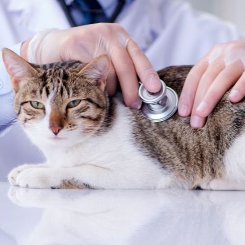
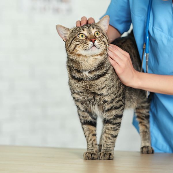
About Our Reidsville Veterinary Hospital
At Reidsville Veterinary Hospital, Inc., we are more than just an animal hospital – we are your dedicated partner in ensuring the well-being of your beloved pets. Our comprehensive services extend beyond routine care to include emergency cases, as well as addressing medical, surgical, and dental needs, whether urgent or less pressing. Our skilled veterinary team is proficient in treating a wide array of conditions and providing various treatments.
As an AAHA-accredited practice, we meet or exceed quality standards set and routinely inspected by the American Animal Hospital Association. We are proud to be an AAHA practice, which helps keep us on the leading edge of veterinary medicine. It also helps us stay focused on continuous improvement to ensure we can offer your pet the quality and range of services you expect and deserve.

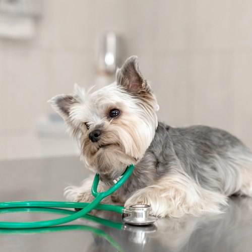
Complete Veterinary Care in Reidsville, NC
At Reidsville Veterinary Hospital, Inc., we treat your pets like the valued family members they are. Every time you visit us, you can expect a relaxed, pleasant atmosphere to help you and your pet feel as comfortable as possible.
Same Day Pet Urgent Care Visits
Pet Preventative and Wellness Care
Full Canine Reproductive Services
Emergency Pet Care
View All Services
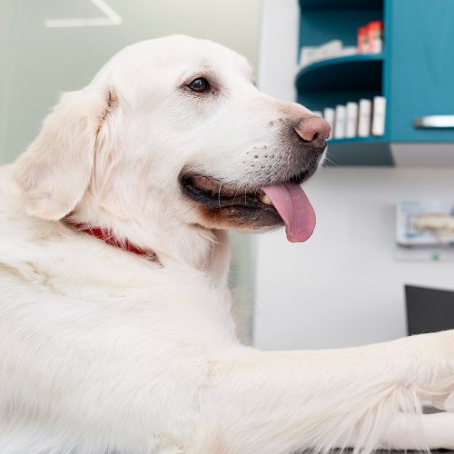
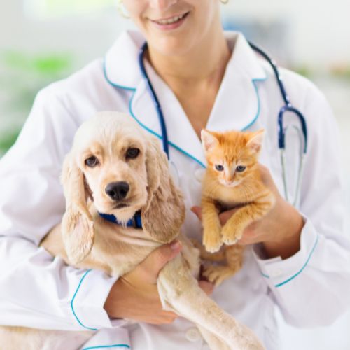
Meet Our Veterinarians and Staff
Our dedicated veterinarians and staff are committed to providing top-notch healthcare for your beloved pets. With years of experience and a genuine love for animals, we ensure your furry friends receive the best treatment possible. From routine check-ups to complex medical cases, we strive to create a warm and welcoming environment where pets and their owners feel comfortable and supported. At Reidsville Veterinary Hospital, your pet’s well-being is our priority, and we look forward to serving you and your four-legged family members with the utmost care and professionalism.